Glossary of Rhinoplasty Terms
Please note that some of the intra-operative photographs contained in this glossary are graphic and may be objectionable or disturbing to some viewers. Use discretion when choosing whether or not to view these educational materials. The images are hidden by a warning message until hovered over with the mouse pointer.
Ala (plural: Alae)
Commonly called the nostril. The paired crescent-shaped convexities flanking the nasal tip that partially surround the nostril openings. The medial aspect of the ala is supported by the lateral crus of the alar cartilage. No cartilage is present in the outer portion of the ala.
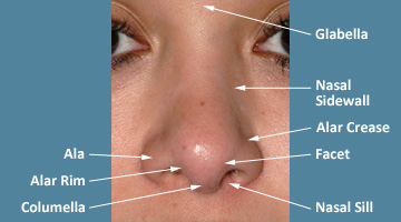 |
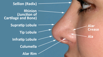 |
| FIGURE 1 Topographic features of the nose; front and profile view. |
Alar Base
The lower-most portion of the nose. Ideal width of the alar base is approximately equal to the inter-canthal distance (distance between the eyes), but may vary according to personal preference. The ideal alar rim configuration is often described as a "gull wing in flight".
 |
FIGURE 2 Alar base width approximates the inter-canthal space, and the nostrils mimic a "gull wing in flight" (shown in red). |
Alar Cartilages
Also called the lower lateral cartilages. Paired (mirror image) arches of flexible nasal cartilage which govern the shape and strength of the columella, nasal tip, and alar rims. Each arch is subdivided into the medial crus (columellar segment), intermediate crus (infra-tip segment), dome (lobular segment or tip-defining point), and lateral crus (alar segment).
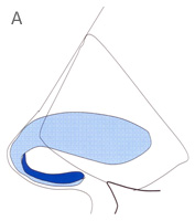 |
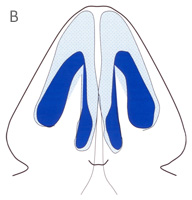 |
| FIGURE 3 Schematic representation of paired alar (tip) cartilages (shown in blue). Profile (A) and base (B) views. | |
Alar Retraction
Also called notched ala or retracted ala. The cephalic mal-position of the alar rim. A common consequence of over-aggressive cephalic trim of the lateral crura, but may also be congenital.
 |
FIGURE 4 Example of severe alar retraction following over-aggressive rhinoplasty. Note excessive nostril show. |
Alar Rim
The outer edge of the nostril opening as seen on front view, and the caudal border of the ala as seen on profile view (cephalad to the columella). Ideally, the alar rim is a gently curved, nearly straight line as seen on profile view. See Figure 1
Autologous Graft
A piece of living tissue (e.g. cartilage, bone, skin, etc.), harvested from the patient’s own body, for the reconstruction of a body part or appendage. In rhinoplasty, cartilage is commonly harvested from the nasal septum and used to reshape or reinforce the nasal tip or nasal bridge. Ear cartilage or rib cartilage may be used when preferable. Cartilage from each donor site has different physical properties, advantages, and disadvantages.
Bony Vault
The upper third of the nose (or upper half of the nasal bridge), composed of the paired nasal bones and the bony septum (vertical ethmoid plate).
 |
| FIGURE 5 Schematic representation of the bony vault (upper nasal third), cartilaginous vault (middle nasal third) and alar cartilage (lower nasal third). |
Bulbous Tip
A cosmetic nasal deformity characterized by a wide oversized nasal tip with large alar cartilage, often having broad rounded alar domes.
 |
FIGURE 6 A typical bulbous nasal tip. Note cupping of the widely spaced and broadly arched alar cartilages seen through thin overlying skin. |
Cartilage Graft
Custom fabricated pieces of cartilage used to strengthen and/or reshape the nasal skeleton. Cartilage grafts may be used to enhance nasal beauty, improve nasal breathing, increase structural stability, or combinations thereof. Cartilage grafts are employed in most forms of contemporary rhinoplasty. See also: Revision Rhinoplasty
 |
FIGURE 7 The non-essential portion of the cartilaginous septum (approximately one inch wide) harvested for graft fabrication. Note the comparatively small amount of cartilage available from the nose. |
Caudal
Synonymous to caudad. Situated nearer to the nasal base (in nasal anatomy only). The opposite of cephalic or cephalad.
Caudal Excess Deformity
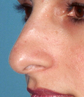
A naturally-occurring cosmetic deformity of the nasal base characterized by caudal over-growth of the nasal septum. Hallmark features are a hanging columella, a short upper lip, and webbing of the nasolabial angle. More commonly seen in Mediterranean and Middle-Eastern noses.
 |
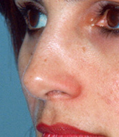 |
| FIGURE 8 Example of a classic caudal excess nasal base deformity before (A) and after (B) rhinoplasty. Note decrease in nasal length and corresponding increase in upper lip length. | |
Cephalic
Synonymous with cephalad (in nasal anatomy only). In a direction toward the head. The opposite of caudal or caudad.
Cephalic Trim
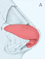
Partial excision of the lateral crus along its cephalic border. A traditional rhinoplasty technique used in tip narrowing and tip rotation. A common cause of alar retraction and other tip deformities.
 |
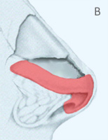 |
| FIGURE 9 Schematic representation of normal alar cartilage (A) and alar cartilage after cephalic trim (B). | |
Closed Rhinoplasty
Also called endonasal rhinoplasty. Refers to type of surgical access to the nasal skeleton associated with a relative lack of surgical exposure. Closed rhinoplasty avoids a visible columellar scar but restricts direct visualization and precludes many techniques currently used in open rhinoplasty. See also: Open vs. Closed Rhinoplasty
 |
FIGURE 10 Closed rhinoplasty in which both alar cartilages are being pulled out ("delivered") through the right nostril for suture modification (i.e., the "delivery" technique). Note the relative concealment of the nasal skeleton, the distortion of the alar cartilages, and the absence of a columellar incision. |
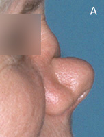
Cocaine Nose
A cosmetic nasal deformity resulting from cocaine abuse and subsequent cocaine-induced tissue necrosis. Hallmark features are a septal perforation, saddle collapse of the middle vault, and progressive deformity. In severe cases, loss of the entire nose and/or fatality may occur.
 |
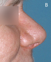 |
| FIGURE 11 Example of advanced cocaine nose deformity before (A) and after (B) surgical reconstruction with rib cartilage. | |
Columella
The central "column" separating the right and left nostrils as seen on base view, composed of skin and the paired right and left medial crura. See Figure 1
Columellar Show
That portion of the columella visible on profile extending beyond the alar rim. Normal columellar show is 2-5 mm.
 |
FIGURE 12 Post-operative example of normal columellar show. Note columellar visibility approximately 3 mm beyond the alar rim. |
Columellar Strut
A specific type of autologous cartilage graft used to increase tip projection, increase tip rotation, and/or enhance tip support. A concealed graft placed within the columella to stabilize the newly shaped nasal tip cartilage and prevent delayed deformity.
 |
FIGURE 13 Schematic showing position of a columellar strut graft (shown in blue) between the right and left alar cartilages. |
Computer Imaging
The process of modifying (i.e. "morphing") a patient’s digital photograph using computer software in order to visually simulate or "preview" various cosmetic changes to the nose for the purpose of clarity. See also: Computer Imaging
Dorsal
Pertaining to the nasal dorsum or nasal bridge (in nasal anatomy only).
Edema
Excessive lymphatic or serous fluid permeation of the soft tissues, typically in response to tissue trauma or disease states. May persist up to 18 months following rhinoplasty in extreme cases. Chronic edema may lead to permanent fibrosis.
Ethnic Rhinoplasty
A term coined to describe cosmetic rhinoplasty in individuals of minority ethnic descent. See also: Ethnic Rhinoplasty
Excisional Rhinoplasty
A term used to describe the traditional method of reduction rhinoplasty in which cartilage excision is the sole means of contour alteration. Although still widely used today, the excisional method is associated with a significantly higher rate of contour and functional deformities. See also: Incisional vs. Excisional Rhinoplasty
Fibrosis
The formation of fibrous (scar) tissue as a reparative or reactive process in organs or tissues not normally containing fibrous tissue. A common sequela in thick-skinned or scar-prone rhinoplasty patients.
Functional Rhinoplasty
A term used to describe surgical reconstruction of the nasal airway to restore nasal function. See also: Functional Rhinoplasty
Hanging Columella
A cosmetic nasal deformity characterized by an overly-protruding columella, resulting in excessive columellar show on profile view.
 |
FIGURE 14 Example of a naturally-occurring "hanging" columella. Note excessive nostril visibility from caudal over-growth of the nasal septum. |
Hypertrophic Scar
An overactive wound healing response to cutaneous injury or surgery, resulting in an unsightly raised and widened scar. Unlike keloids, a hypertrophic scar usually responds to timely treatment.
Inferior
Situated toward the bottom or below. Situated nearer the soles of the feet in relation to a specified reference point. The opposite of superior.
Infra-tip Lobule
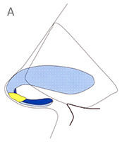
The lower-most portion of the nasal tip found immediately above the nostril openings (See Figure 1). The infra-tip lobule is formed by the paired (divergent) intermediate crura of the alar cartilages. See Figure 15
Intermediate Crus (plural: Crura)
The segment of alar cartilage found above the nostril and below the nasal dome, corresponding to the infra-tip lobule.
 |
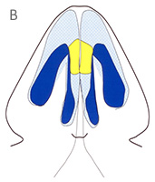 |
| FIGURE 15 Schematic of the intermediate crura (shown in yellow) from the profile (A) and base (B) views. | |
Intermediate Osteotomy
A modified traditional bone cut positioned midway between the medial and lateral osteotomy cut. Used to reshape misshapen, indented, or cupped nasal bones.
Inverted-V Deformity
A nasal bridge shadow deformity resulting from excessive narrowing of the middle vault cartilage relative to the adjacent nasal bones. A type of step-off deformity resulting from over-aggressive nasal hump reduction or blunt nasal trauma.
 |
FIGURE 16 Example of inverted-V deformity. Note inverted V-shaped shadow formed by middle vault pinching after over-aggressive hump reduction. |
Keloid
A severe overactive wound healing response to cutaneous injury or surgery, resulting in a stubborn tumor-like accumulation of fibrous (scar) tissue. Often difficult or impossible to treat in susceptible individuals.
Kenalog
The brand name of triamcinolone acetate, an injectable synthetic steroid used to prevent prolonged edema and unwanted fibrous (scar tissue) formation.
Lateral
Farther from the midline or mid-sagittal plane. Toward the outer perimeter. The opposite of medial.
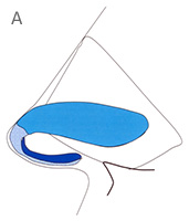
Lateral Crus (plural: Crura)
A sub-component of the alar (tip) cartilage. The lateral crus extends obliquely immediately above the alar crease from the nasal dome (medially) to the pyriform aperture/cheek (laterally).
 |
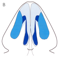 |
| FIGURE 17 Schematic of the lateral crura (shown in turquoise) from the profile (A) and base (B) views. | |
Medial
Closer to the midline or mid-saggital plane. Near the middle. The opposite of lateral.
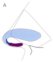
Medial Crus (plural: Crura)
The caudal/inferior most segment of the alar cartilage corresponding to the columellar segment.
 |
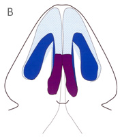 |
| FIGURE 19 Schematic of the medial crura (shown in purple) from the profile (A) and base (B) views. | |
Medial Osteotomy
A para-sagittal bone cut, placed immediately adjacent to the sagittal midline, for medial repositioning (infracture) of wide nasal bones. Used in tandem with lateral osteotomy for complete infracture. See Figure 18
Middle Vault
The middle third of the nose (and the lower half of the nasal bridge). Composed entirely of cartilage, the middle vault is formed by the mid-line dorsal septum and the right and left upper lateral (sidewall) cartilages. See Figure 5
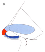
Nasal Domes
The hinge-point (apex) of each alar cartilage formed by the junction of the intermediate and lateral crus. The tip-defining point.
 |
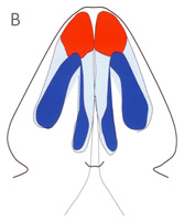 |
| FIGURE 20 Schematic of the nasal domes (shown in red) from the profile (A) and base (B) views. | |
Nasal Dorsum
Synonymous with nasal bridge. The upper two-thirds of the nose consisting of the middle (cartilaginous) vault and the upper (bony) vault.
Nasal Implant
A non-living, synthetic material used to replace missing or damaged nasal tissues. Although sometimes controversial, nasal implants are still used routinely.
Nasal Tip
Also called the lobule. The forward-most part of the nose, formed by the alar domes, flanked on either side by the alae. Subdivided into the tip lobule, supra-tip lobule, and infra-tip lobule. See Figure 1
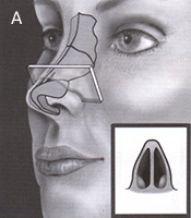
Nasal Valve
The natural anatomic "bottleneck" of the nasal airway which regulates nasal airflow through alternate swelling and contraction of the septal and turbinate mucosa found on its inner surface. Anatomic obstructions to nasal airflow are most common in the nasal valve area due to its naturally small size. Cosmetic nasal surgery is a common cause of nasal valve dysfunction.
 |
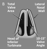 |
| FIGURE 21 Schematic cross-section of the nose taken through the nasal valve area (A) demonstrating the small cross-sectional area and partial obstruction by the inferior turbinate head (B). | |
Nasofrontal Angle
The angle formed between the nasal bridge and the forehead (glabella) as seen on profile view. Ideally the apex (or nasal starting point) is situated at the level of the superior eyelash line.
Nasolabial Angle
The angle formed between the columella and the upper lip as seen on profile. Typically more obtuse in females. See Figure 22
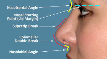 |
| FIGURE 22 Profile aesthetics of the nose. |
Necrosis
Tissue death resulting from inadequate oxygenation, usually a result of disrupted blood flow.
Open Rhinoplasty
Open, or external, rhinoplasty refers to a more aggressive surgical exposure of the nasal skeleton. With the aid of a small but visible skin incision on the columella, open rhinoplasty offers direct visualization of the nasal skeleton. Most experts regard open rhinoplasty as more versatile, and the exposure of choice for complex anatomy or revision rhinoplasty. In expert hands, the columellar scar is often invisible. See also: Open vs. Closed Rhinoplasty
 |
FIGURE 23A Incision placement for open rhinoplasty. Note the V-shaped trans-columellar incision extending inside the nostril. |
 |
| FIGURE 23B Typical skeletal exposure obtained using open rhinoplasty. Note direct visualization of the undistorted nasal framework. |
Open Roof Deformity
Gapping of the nasal bones following dorsal hump reduction resulting from over-aggressive hump reduction and/or failure to completely infracture the nasal bones.
Percutaneous Osteotomy
Also called postage-stamp osteotomy. A type of lateral osteotomy employing a series of side-by-side bone cuts made through a single small skin incision. Thought to be less difficult than the traditional (endonasal) lateral osteotomy, but with the potential for a visible scar.
Pinched Lobule
Excessive narrowing of the nasal tip secondary to medial collapse of the alar domes. A frequent consequence of over-aggressive alar cartilage removal and/or over-aggressive tip sutures.
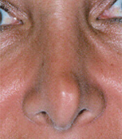 |
FIGURE 24 Example of pinched lobule (tip). Note unsightly vertical flattening of the nasal tip. |
Primary Rhinoplasty
Primary rhinoplasty refers to a previously unoperated nose or first-time rhinoplasty. Primary rhinoplasty is not hindered by previous surgical scarring of the soft tissues or previous surgical damage to the nasal cartilage or bone.
Radix
The nasal root. The upper termination of the nasal bridge at the glabellar base. A soft tissue landmark which roughly corresponds to the deepest point of the nasal bone (i.e., the nasion). See Figure 1
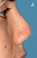
Retracted Columella
A columellar deformity characterized by little or no columellar show, resulting from inadequate columellar projection secondary to over-aggressive cosmetic surgery, nasal trauma, or birth deformity.
 |
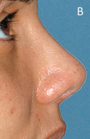 |
| FIGURE 25 Example of retracted columella with minimal columellar show before (A) and after (B) correction. | |
Rhinion
Junction of the bony upper nasal vault with the cartilaginous middle nasal vault. The typical high point of a nasal bridge hump. See Figure 1
Rotation
Term used to describe tip position along a fixed arc of rotation. A "rotated tip" rests closer to the forehead and appears perky, whereas a counter-rotated tip is positioned away from the forehead and may appear droopy. Ideal tip rotation varies according to personal preference.
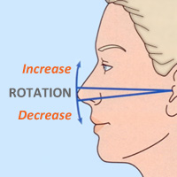 |
FIGURE 26 Schematic representation of tip rotation. |
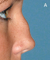
Saddle Nose Deformity
A cosmetic and functional nasal deformity resulting from collapse of the middle vault. Causes include autoimmune disease, cocaine abuse, blunt trauma, and over-aggressive cosmetic nasal surgery.
 |
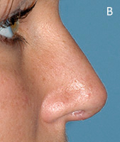 |
| FIGURE 27 Example of auto-immune-induced saddle nose deformity before (A) and after (B) rib graft reconstruction. | |
Secondary Rhinoplasty
Secondary or revision rhinoplasty refers to re-operation of a previously operated nose. Secondary rhinoplasty may be performed for cosmetic and/or functional enhancement. Revision rhinoplasty is generally regarded as one of cosmetic surgery’s most difficult operations owing to pre-existing skeletal damage and soft tissue scarring. In general, the recovery is slower and the outcome less dramatic relative to a well-executed primary rhinoplasty. See also: Revision Rhinoplasty
Septum
The dividing wall separating the right and left nasal passages, formed by cartilage or bone and covered on both sides with nasal mucosa.
Septal Extension Graft
A type of columellar strut graft attached to the caudal septum for maximum support. Commonly used to lengthen short noses.
Shield Graft
A type of infra-tip cartilage graft fixed to the alar cartilages in order to counter-rotate and/or increase tip projection.
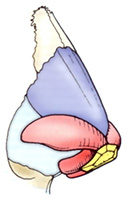 |
FIGURE 28 Schematic representation of a shield-type augmentation graft (shown in yellow) placed in the infra-tip lobule. |
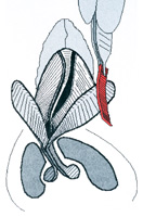
Spreader Graft
A middle vault augmentation graft placed between the dorsal septum and the upper lateral cartilage(s) to increase middle vault width, improve symmetry, increase airway dimension, and/or straighten the dorsal septum.
 |
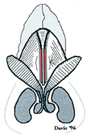 |
| FIGURE 29 Schematic representation of bilateral spreader graft (shown in red). Placement of graft in middle vault straightens curvature of the dorsal septum. | |
Structural Rhinoplasty
A philosophical approach to both cosmetic and
functional rhinoplasty, in which nasal framework is artfully reconfigured
to enhance beauty, and then structurally reinforced to ensure lasting
results. The philosophical opposite to excisional rhinoplasty,
a traditional approach in which structural support is compromised, leading
to a high probability of progressive deformity.
See
also: Incisional
vs. Excisional Rhinoplasty
Superior
Situated toward the top or above. Situated nearer to the vertex (top of the head). The opposite of inferior.
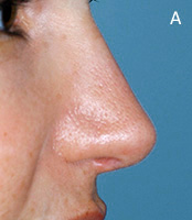
Supra-Tip Break
Also called retroussé (pronounced reh-troo-SAY). A naturally-occurring variation in height between the nasal tip and the adjacent nasal bridge (as seen on profile). A common topographic feature in feminine noses often sought in cosmetic nasal surgery. The ideal tip retroussé varies according to personal preference and can be modified accordingly.
 |
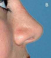 |
| FIGURE 30 Examples of post-operative supra-tip retroussé, ranging from mild (A) to pronounced (B). | |
Tension-Nose Deformity
A naturally-occurring cosmetic deformity characterized by dorsal overgrowth of the nasal septum. Hallmark features include "pollybeak" overgrowth deformity of the nasal bridge, overprojection of the nasal tip, and a wide nasal pedestal.
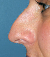 |
FIGURE 31 Example of a classic tension nose deformity, as seen on profile view. Note overprojected tip and pollybeak deformity (overprojected middle vault). |
Tip Projection
Term used to denote horizontal protrusion of the nose from the vertical plane of the face.
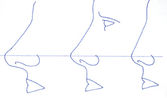 |
| FIGURE 32 Schematic representation of tip projection, including over-projected (left), normal (middle), and underprojected (right). |
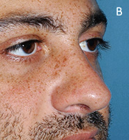
Tongue-in-Groove Setback
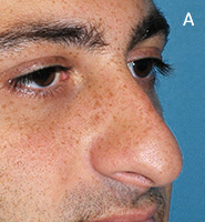
A versatile surgical technique used to treat the caudal excess nasal deformity by sewing the medial crura to the caudal septum.
 |
 |
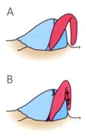 |
| FIGURE 33 Example of caudal excess nasal deformity (A), corrected with tongue-in-groove setback procedure (B). | ||
Turbinate
Mucosa-covered bones spanning the length of both nasal passages responsible for warming, filtering, and humidifying the inspired air; and for regulation of nasal airflow. Turbinate overgrowth (hypertrophy) is a common cause of nasal airway obstruction. See also: Functional Rhinoplasty
